


This article will review the material you have learned in A&P class. Knowing and understanding the main anatomy and organ functions of each human body system will set you on track towards scoring higher on the HESI without studying harder or spending hours poring over every detail of your massive textbook. As you come closer to completing your prerequisites, it is now time to begin preparations to take the HESI A2 Entrance Exam in order to apply and get into the nursing school of your choice.
The HESI A2 Entrance Exam is an assessment for schools to know if you are ready to start your education in your chosen health career. This is why the HESI is used by other healthcare programs and not just nursing schools. Students who are studying to become dental hygienists, radiology techs, etc., must also take the HESI exam to move forward with their program. Each program has different requirements, but one section of the HESI that is guaranteed to be mandatory for nursing schools is the HESI Anatomy and Physiology section.
Like many students, you have probably spent the last two years staying up late studying for your various nursing prerequisite classes. In fact, you may have even pulled an all-nighter or two, studying with friends before an anatomy exam. This approach can be physically and mentally exhausting and can potentially impact your testing performance. Often, students show up to an exam after countless hours of preparation only to discover they spent too much time studying some concepts and not enough on others. Other students who are balancing jobs, family, and other obligations have difficulty finding time to study at all!
But do not panic just yet! This article serves as a HESI Anatomy and Physiology study guide and will demonstrate that it is not necessary to devote hours and hours of your studying time to comb over every anatomical function or organ of the body.
For example, you’ll find that knowing which hormones are produced by the anterior pituitary gland and which hormones are produced by the posterior pituitary gland is a bit too specific for the HESI. However, grasping which hormones are produced by the pituitary gland in general, is an effective strategy for test preparation, as it will help you grasp the necessary material and it will save you time and brainpower!
HESI Anatomy and Physiology Study Guide
It is highly probable that if you put a significant amount of your time and energy towards memorizing and internalizing the key terms in this article, you can do well without even cracking open your huge 1000-page anatomy textbook. As long as you know the basics and general terms of each human body system, rest assured that you WILL pass the anatomy and physiology section. However, this doesn’t mean you shouldn’t study at all. Make sure to go over your study guide and practice questions.
So, let’s get started!
1. Body Planes
Planes of the body are critical to know as you begin your A&P studies because they provide important reference points throughout the human body.
The three planes of the body are the frontal (coronal), transverse, and sagittal planes.
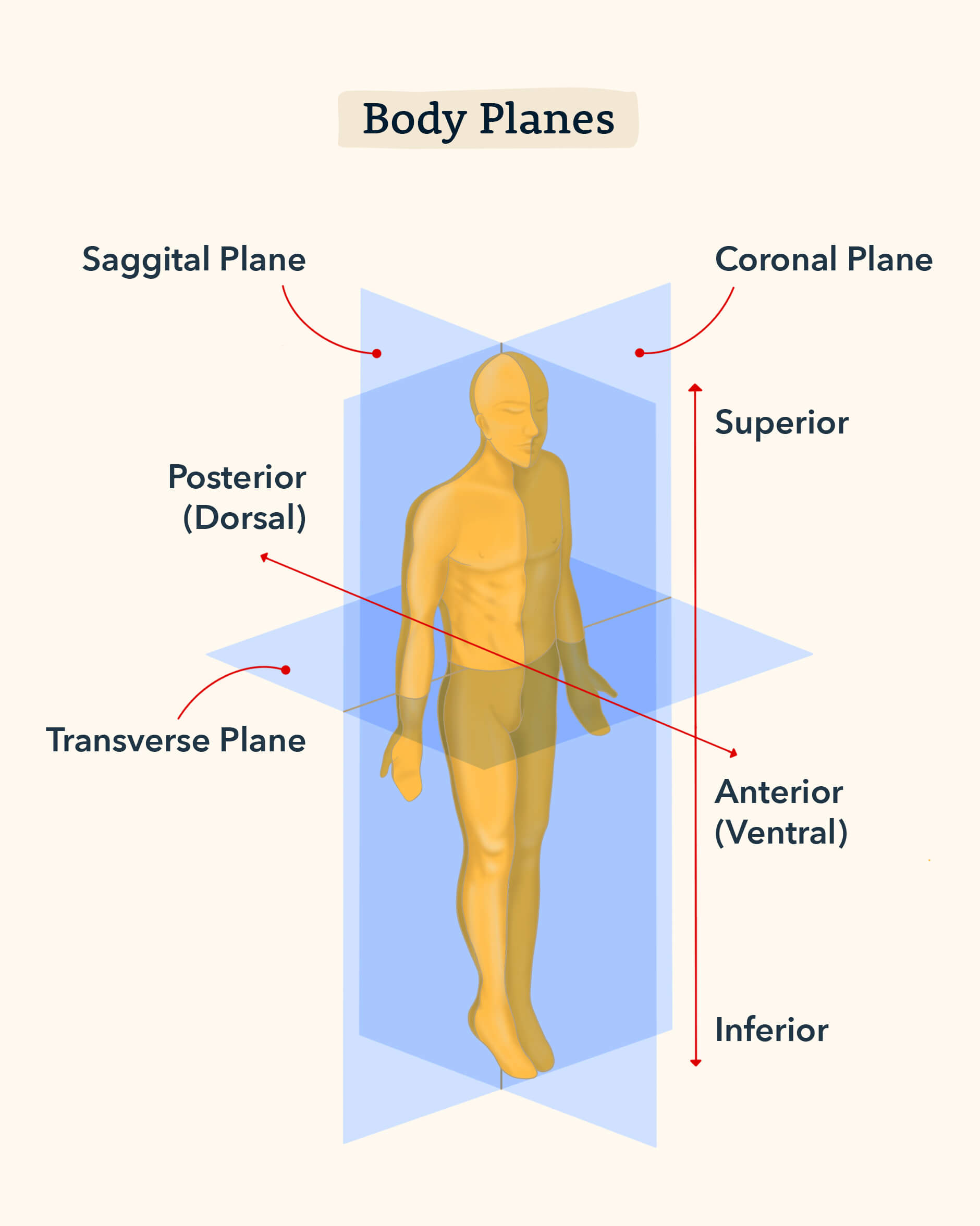
Sagittal Plane
Cuts or divides the body into right and left portions. Always associate the sagittal plane with your right and left side, like the right and left turning signals of a car. When a driver wants to signal a right turn, the right blinker turns on and the left blinker remains off. Alternatively, when a left signal is needed, only the left blinker should be activated.
Coronal (Frontal) Plane
Cuts or divides the body into front (anterior) and back (posterior) portions. Imagine standing in line at the grocery store. People stand behind one another at a frontal plane.
Transverse (Horizontal) Plane
Cuts or divides the body into top (superior) and bottom (inferior) portions. Think of the sun setting over a body of water: you have the sun at the top, the ocean at the bottom, and the transverse plane (horizon) in the middle. In this way, a sunset creates a transverse plane.
2. Body Orientation and Terms of Direction
You will find that once you are able to distinguish between body planes, it is significantly easier to describe body parts according to their orientation and direction. Note that these keywords are almost always asked on the HESI, so be sure to memorize them!
- Superior: Above
- Inferior: Below
- Anterior (Ventral): Towards or in front of the body
- Posterior (Dorsal): Behind, towards the backside of the body
- Medial: Closer to the midline of the body
- Lateral: Farther away from the midline of the body
- Proximal: Closer or toward the body trunk/chest/center of the body
- Distal: Farther and distancing away from the body trunk/chest/center of the body
- Superficial: Closer to the outer layer and surface of the body
- Deep: Away from the surface of the body
As a HESI test taker, it is imperative to be able to correctly use the directional terms and their definitions, interchangeably. For example, if you are asked a question about the Respiratory System, how would you answer the following question:
What is anterior to the esophagus?
A. Trachea
B. Lungs
C. Bronchioles
D. Larynx
So, in order to answer this question, you’ll need to consider the definition of anterior. What is anterior (in front) of the esophagus?
Your answer should be that the trachea is in front of the esophagus. Ensure that you are particularly comfortable with sentences like the following:
- The neck is inferior to the face.
- The chin is superior to the navel.
- The rib cage is ventral to the heart.
- The nose is medial to the eyes.
- The ears are lateral to the eyes.
- The fingers are distal to the wrist.
- The wrist is proximal to the fingers.
And what helps us move all these parts and sections of our bodies? Bones!
3. The Skeletal System
For the skeletal system, the HESI tends to test moreover anatomical questions than physiology. However, let’s cover a few physiological points before we review the anatomy.
-
- Osteogenic Cells: are the only bone cells that divide and become osteoblasts
- Osteoblasts: are cells that secrete matrix for bone formation
- Osteocytes: are bone cells embedded in the matrix
- The Haversian Canal: are tiny tubes that form a network to communicate with bones and their blood vessels
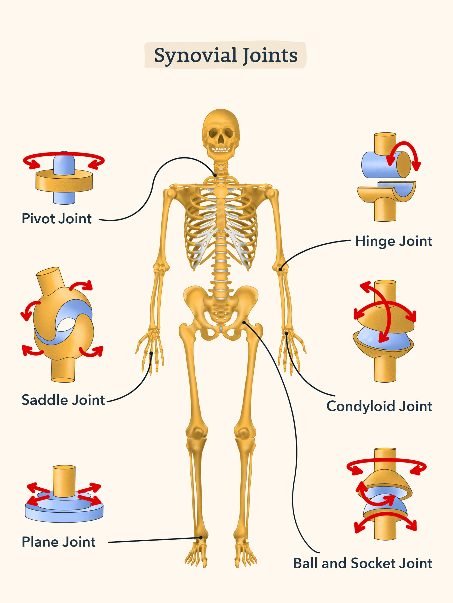
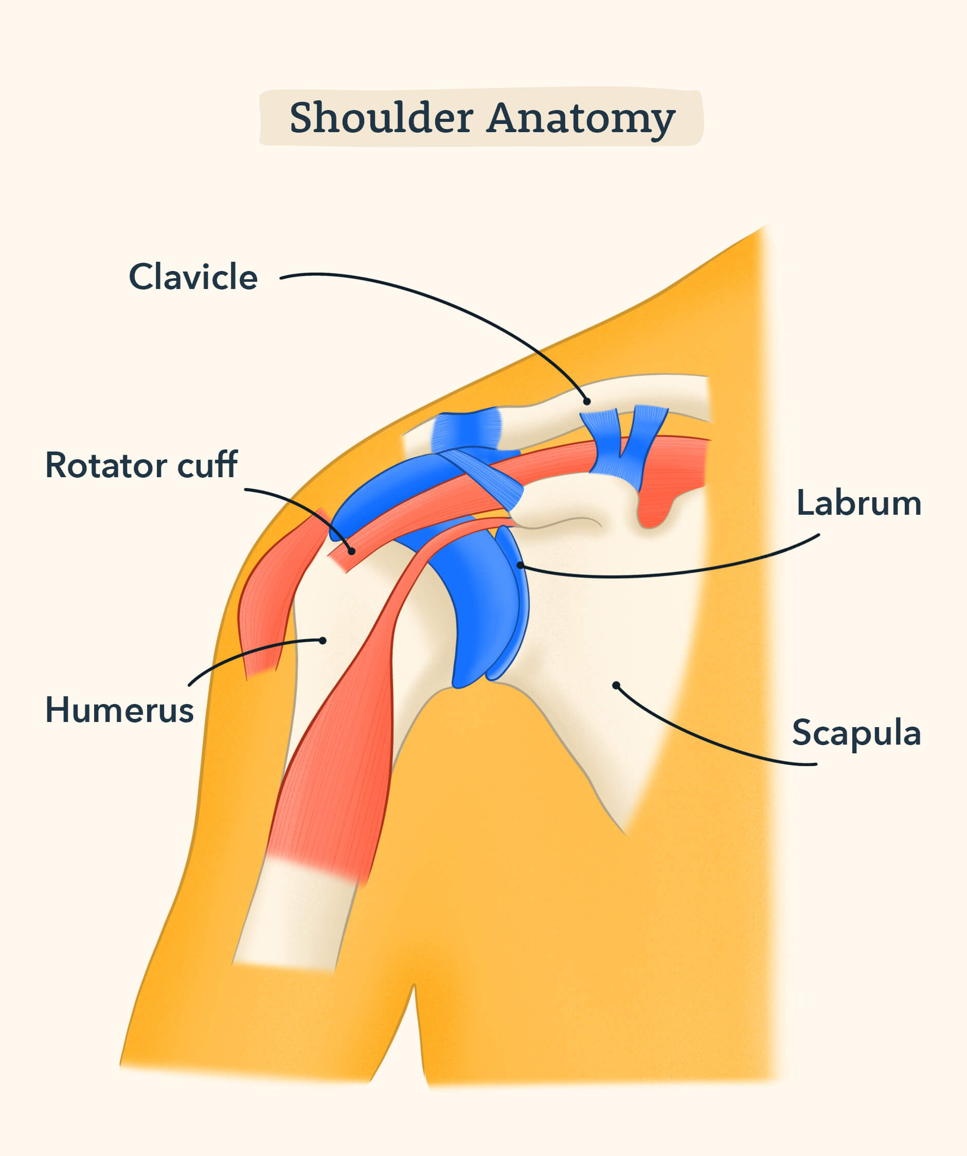
- Types of Synovial Joints: ball and socket (shoulder and hip), hinge (elbow and knee)
Did you know the average human adult has 206 bones? Yikes! Luckily, you do not need to memorize all of them for your HESI exam. The major bones of the body are as follows:
- Head: parietal and frontal bone
- Chest: scapula, clavicle, ribs, sternum
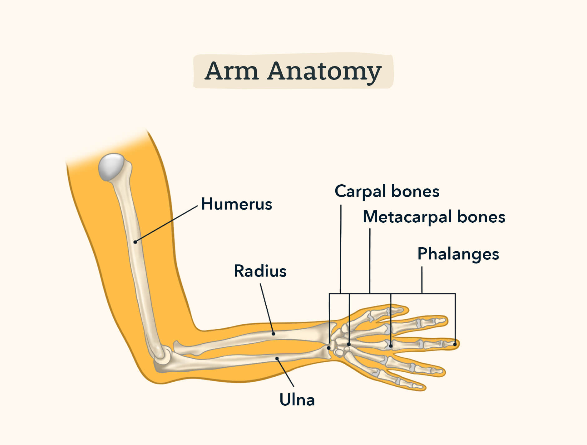
- Arm: humerus, radius, ulna
- Leg: tibia, fibula, femur
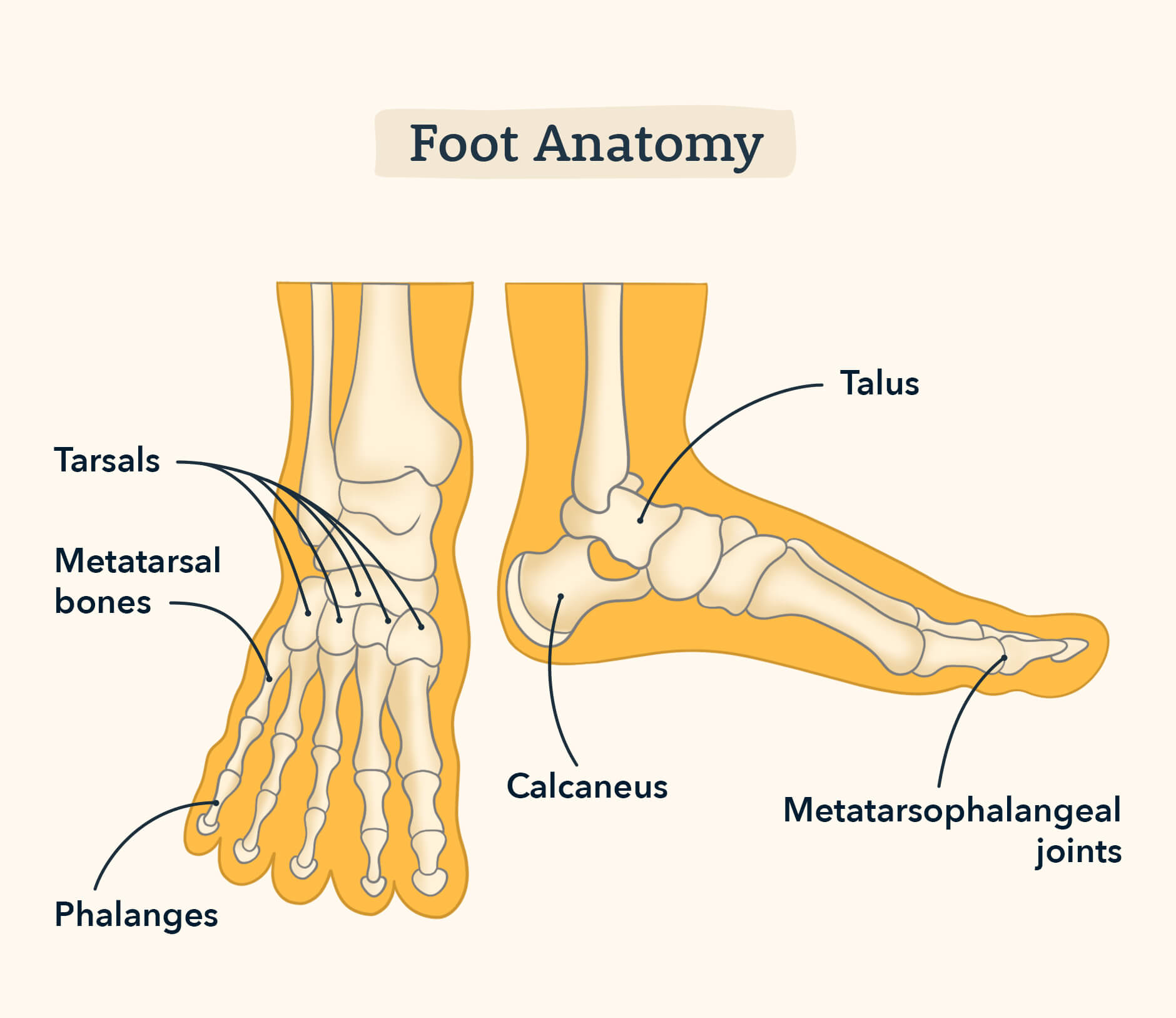
- Feet: tarsals (heel), metatarsals (midfoot), phalanges (toes)
And can you guess what is wrapped around our bones to allow protection, movement, and strength? Muscles!
4. The Muscular System
When we go to the gym, we usually try to focus on some specific areas when lifting weights: abs, biceps, triceps, etc. But there’s a whole lot more going on in our muscular system than just those muscles you’ve been trying to build in the weight room.
In regard to preparing for HESI questions related to the muscular system on your exam, your best bet is to focus on the anatomy rather than the physiological aspects of the muscular system just as in the case of the skeletal system.
Before we get into the muscles, let’s take a minute to look at the three layers of connective tissue:
- Epimysium: Outermost layer
- Perimysium: Surrounds muscles fibers
- Endomysium: Deepest layer of muscles
Note that the major regional muscles that are common in the HESI anatomy and physiology section are included in the list below.
Face
- Temporalis (side of head)
- Zygomaticus (cheek area)
Leg
- Quadriceps (rectus femoris, vastus lateralis, vastus medialis, vastus intermedius)
- Gastrocnemius (the calf area)
Chest
- Deltoid (shoulder area)
- Pectoralis major and minor (chest, near breastbone)
- Serratus anterior (near the upper ribs)
There is one muscle (muscle organ) we did not mention earlier…and not because it’s not important. On the contrary, it is a muscle that we need more than any other muscle in order to survive. Did you guess? It is the heart!
5. The Circulatory System
If there is one system you should master from this entire article, it is this one. Perhaps more than any other section, this is the section you will most benefit from reading attentively and carefully. Heck, print this part now!
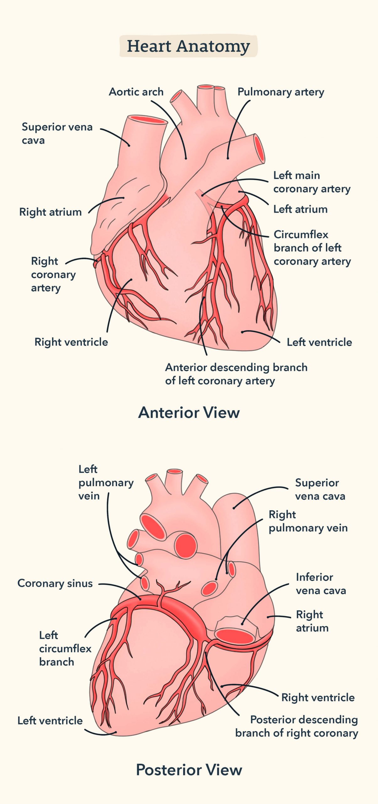
The heart flow is critical to know. You must know the correct order in which the blood enters the heart and goes back out to the rest of the body.
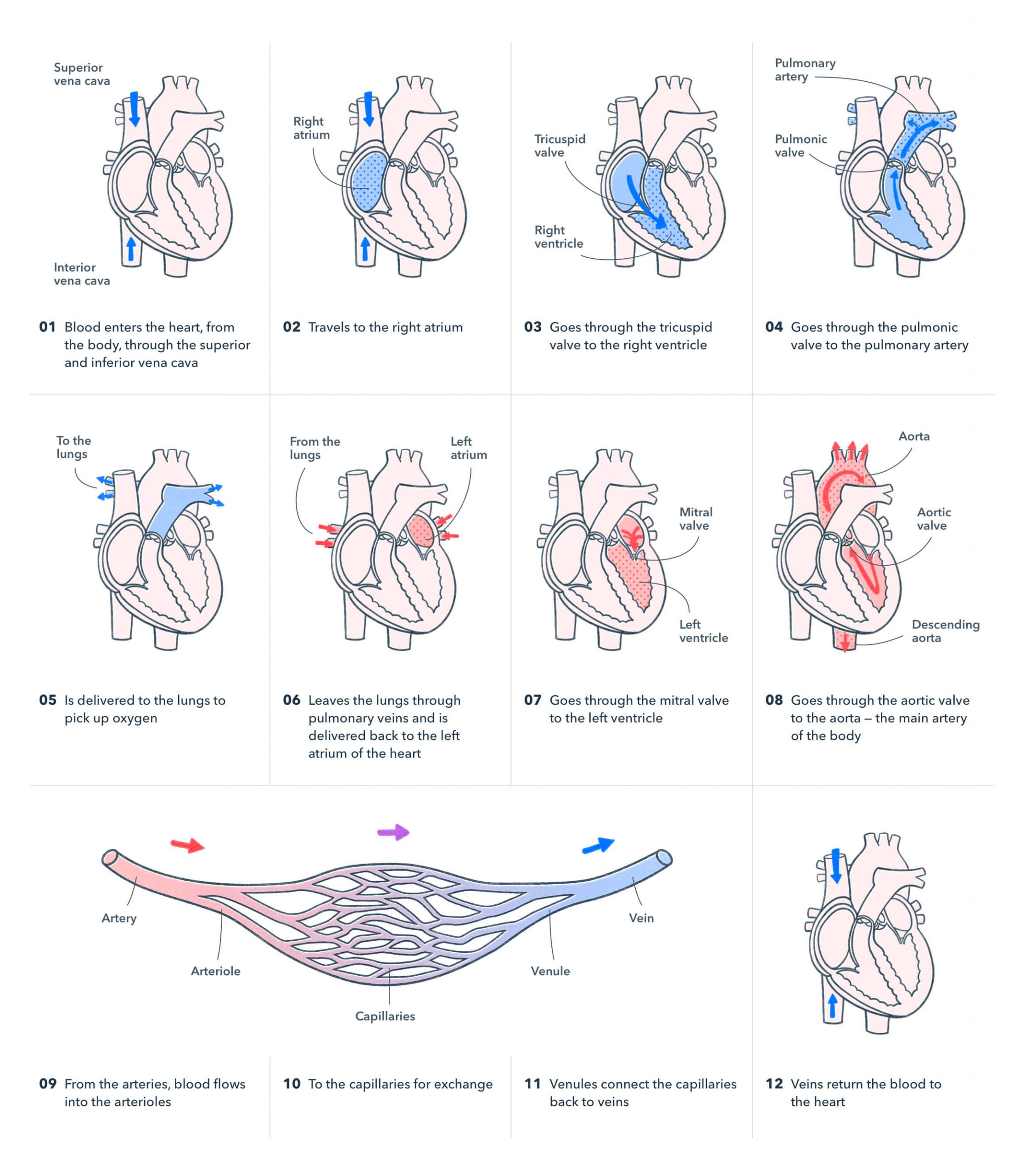
There are two kinds of blood flows in the complex organ of the heart. One flow takes deoxygenated blood AWAY FROM our body to the heart and the other flow carries fresh, oxygenated blood TO the body from the heart.
Both flows will always go from the VEINS ATRIUM VALVE VENTRICLE VALVE again and then finally out of the ARTERY.
Remember: We start with veins. We end with arteries.
The tricky part to remember is that deoxygenated flow will start from the right side and the oxygenated fresh flow will start from the left side of the heart.
For the left side, it might be useful to remember the phrase “fresh to left,” in reference to the popular slang phrase “fresh to death,” meaning something so good, it is inexplicable and what’s better than freshly oxygenated blood to our bodies!
Okay so now you’re thinking, “Fine, I get it. Left is oxygenated and right is deoxygenated, but how will I remember the valves? What if I confuse them?”
First off, remember that valves are like doors, they must be opened before you go inside. So a valve (or door) will need to be opened before the blood moves from the atrium to the ventricle. There will always be a door (valve) between the atrium and ventricle, whether it is the right or left side.
The same thing is true for the ventricle and artery. There will always be a door (valve) that needs to open before the blood moves from the ventricle to the artery.
Now let’s take that slang phrase to another notch, “fresh 2 left.” The left oxygenated side has the bi (two) cuspid valve. So when you think of “fresh 2 Left,” remember “fresh (new oxygenated blood) 2 (bicuspid valve) Left.”
You may also find it helpful to remember that another name for the bicuspid valve is the mitral valve.
Oxygenated blood will flow from the pulmonary (think of lungs and fresh oxygen) veins to the Left Atrium. Then, the bicuspid or mitral valve door is opened so the blood can flow into the Left ventricle. Finally, the aortic valve door is opened so the fresh blood can rush out of the aorta and into the rest of our bodies.
The same exact flow happens for deoxygenated blood except, it’s on the right side of our heart:
This water flow of blood will rush in through the vena cava, drop into the right atrium, and as the blood starts to fill, the tricuspid (also known as AV) valve will open to let the blood flow lower into the right ventricle. The blood needs to leave the ventricle so when it starts filling up, the pulmonary valve gets the cue to open, and then the blood travels to the pulmonary artery.
One tip is to write this down as soon as you are at the testing center, in front of your computer, before you even begin your first section. You will be given sheets of scratch paper, so write this and any other information you can.
Next, we will cover three more must-know topics in relation to the Circulatory System and main points that you must know for the HESI A2 Exam.
The adult human heart has three layers of heart muscles. They include the following:
- Epicardium: The outermost layer and is in contact with the serous connective layer called the pericardium
- Myocardium: The second layer of the heart including the cardiac walls which lets the blood flows in and out
- Endocardium: Made up of the simple squamous epithelium cells and lines the inside of the heart chambers and the surface of the valves
These intricate features of the heart help us survive and flourish on a day-to-day basis. Whether we’re just sitting around reading a book or exercising strenuously, the blood flow through our heart helps the rest of our body receive oxygen.
Now that you understand the “heart of the matter” when it comes to our circulatory system, let’s get to know our immune system.
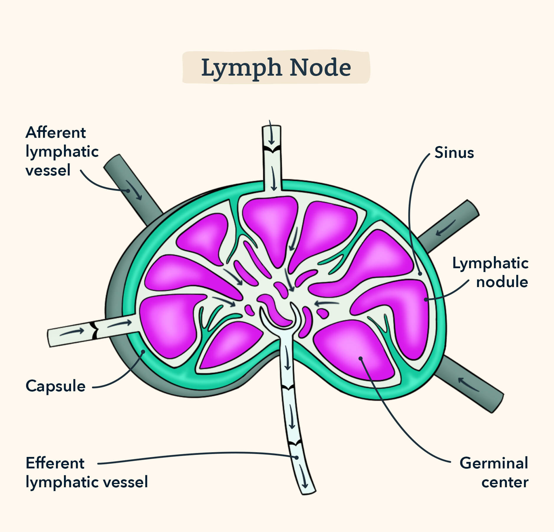
6. Immune/Lymphatic System
When you go to see a doctor, you’ll notice that in addition to taking your temperature, heart rate, and blood pressure, they will usually gently press your neck just below your jawline in a circular motion. Did you ever wonder what this is for? They’re checking for swollen lymph nodes, an indicator of sickness.
The key physiological terms of the immune system are the following:
- T cells: lymphocytes (white blood cells) are produced in the bone marrow, then move into the thymus, which is what the “t” stands for
- B cells: lymphocytes which are produced in the bone marrow and are the surveillance for pathogens
- Thymus: an organ which lies behind the sternum and in front of the trachea, t cells are stored here and is at its largest size during childhood
- Basophil: white blood cells with granulocytes, least common of all other granulocytes
- Eosinophil: white blood cells which are “acidic loving” granulocytes
- Neutrophil: white blood cells with granulocytes and most abundant in our blood
- Plasma/Thrombocytes: platelets and cell fragments that do not have a nucleus but help in creating blood clotsz
And just like that, we wrap up the immune system. There is not as much anatomy to memorize, but understanding the function of this human body system will help you succeed with any immunity-related question.
7. The Endocrine System
The hormones produced by the glands in the endocrine system are released into the blood, depending on what is happening to our body and how our body needs to react. This includes body functions like metabolism, temperature, moods, growth, and development.
The following is a list of major hormones of the endocrine system:
- Antidiuretic hormone (ADH): is secreted by the posterior pituitary gland and acts on the kidney to preserve fluid and electrolyte balance by increasing water reabsorption
- Luteinizing hormone (LH): is secreted by the anterior pituitary gland and occurs midway through the menstrual cycle, it triggers ovulation and creates the corpus luteum
- Follicle stimulation hormone (FSH): is secreted by the anterior pituitary gland and helps eggs mature and causes the menstrual cycle to start in females at puberty.
- Prolactin: is secreted by the anterior pituitary gland and is responsible for milk production; lactation
- Estrogen: is produced in the sex organs and promotes growth and development in females
- Testosterone: is produced in the sex organs and promotes growth and development in males
- Aldosterone: is produced by the adrenal gland and increases reabsorption of sodium ions (and eventually water like ADH) from the nephron
- Oxytocin: is secreted by the posterior pituitary gland and triggers childbirth
- Serotonin: is the main hormone for regulating mood and feelings of well-being
The major glands of the endocrine system produce the above-mentioned hormones.
- Pituitary gland: is at the base of the brain, right under the hypothalamus, and is considered the “master” gland because it affects other glands in the body
- Pineal gland: is situated right between the two brain hemispheres and produces the hormone melatonin which modulates sleep patterns
- Adrenal gland: is found on top of the kidney and secretes cortisol
- Parathyroid gland: regulates the amount of calcium that flows into the blood and bones
- Hypothalamus: an organ in the brain that is responsible for keeping our body at homeostasis, it is considered the “command center’ of the brain” and is anatomically attached to the pituitary gland and therefore controls body temp., fatigue, hunger, and thirst.
8. The Nervous System
The nervous system consists of the anatomy of the brain and the communication between neurons and other parts of the human body.
A good way to compare the nervous system is to the electric wiring of a home. The home is your body and the electric wiring is like your nervous system communicating within walls and knowing where to turn on the light in a specific part of the house.
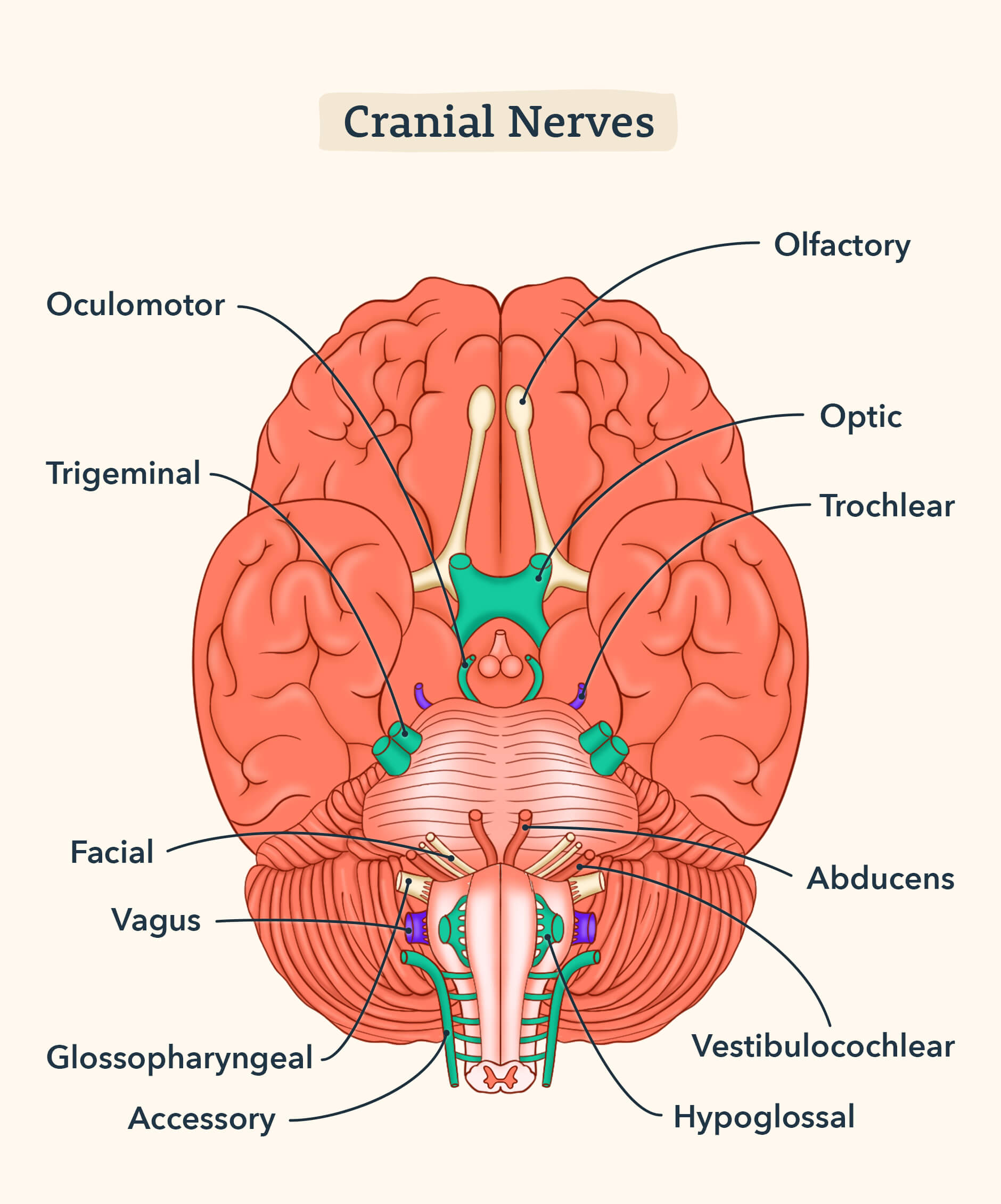
Our nervous system is divided into two parts: the central nervous (brain and spinal cord) and the peripheral nervous system (the cranial nerves and neurons in our body).
The peripheral nervous system receives sensory information from our body, the outside world, external stimuli and then relays that information to the central nervous system.
The peripheral nervous also has two systems within itself:
- Parasympathetic system: this system helps the body and muscles while the vagus nerve slows down the heart rate, “rest and digest” response
- Sympathetic system: this system helps the body react quickly, muscles are tense, “fight or flight” response
Check out this video for a further explanation of the parasympathetic and sympathetic system.
Be sure to memorize and understand the following anatomical and physiological terms of the nervous system:
- Cerebellum: directs motor control (muscle coordination), balance, and equilibrium
- Cerebrum: assists with motor control and cognitive functions such as learning
- Medulla oblongata: brain stem of the brain which connects the spinal cord to the brain; controls autonomic functions (parasympathetic, sympathetic)
- Olfactory nerve: sensory component for the sense of smell
- Optic nerve: registers visual information
- Vagus nerve: slows down the heart rate
9. Body Senses
Think about the senses you use every day. You walk by the local pizza parlor one afternoon and the delicious smell has you craving a slice. You walk in and the pizza maker hears the bell on the door ring, so he is now aware a customer has arrived. He takes your order and cuts you a slice of pepperoni. Before handing it over to you, he warns you that the pizza just came out of the oven, so it’s hot. But you’re so hungry you can’t help yourself. You take a bite and—ouch! He wasn’t kidding! You’ve burnt the roof of your mouth with the hot cheese! But the pizza still tastes just as delicious as it smelled. Maybe you’ll even order another slice!
Think about how you used each of your senses in this scenario and how they influenced you.
Now we will learn a bit more about how these senses work. The body senses that we use every day involve the following organs:
Ear
- Hearing and equilibrium: controlled by the semicircular canals
- Anatomy of the ear: auricle, external auditory meatus
- Cochlea: sends sounds to neurons
- Middle ear: sends sounds from the outer ear to the inner ear
- Eardrum: also called tympanic membrane, vibrates with sound from the outside to travel into the middle ear
Nose
- Olfactory nerve: transmits odors
- Olfactory organ: function for smelling, and warming/filter air we breathe
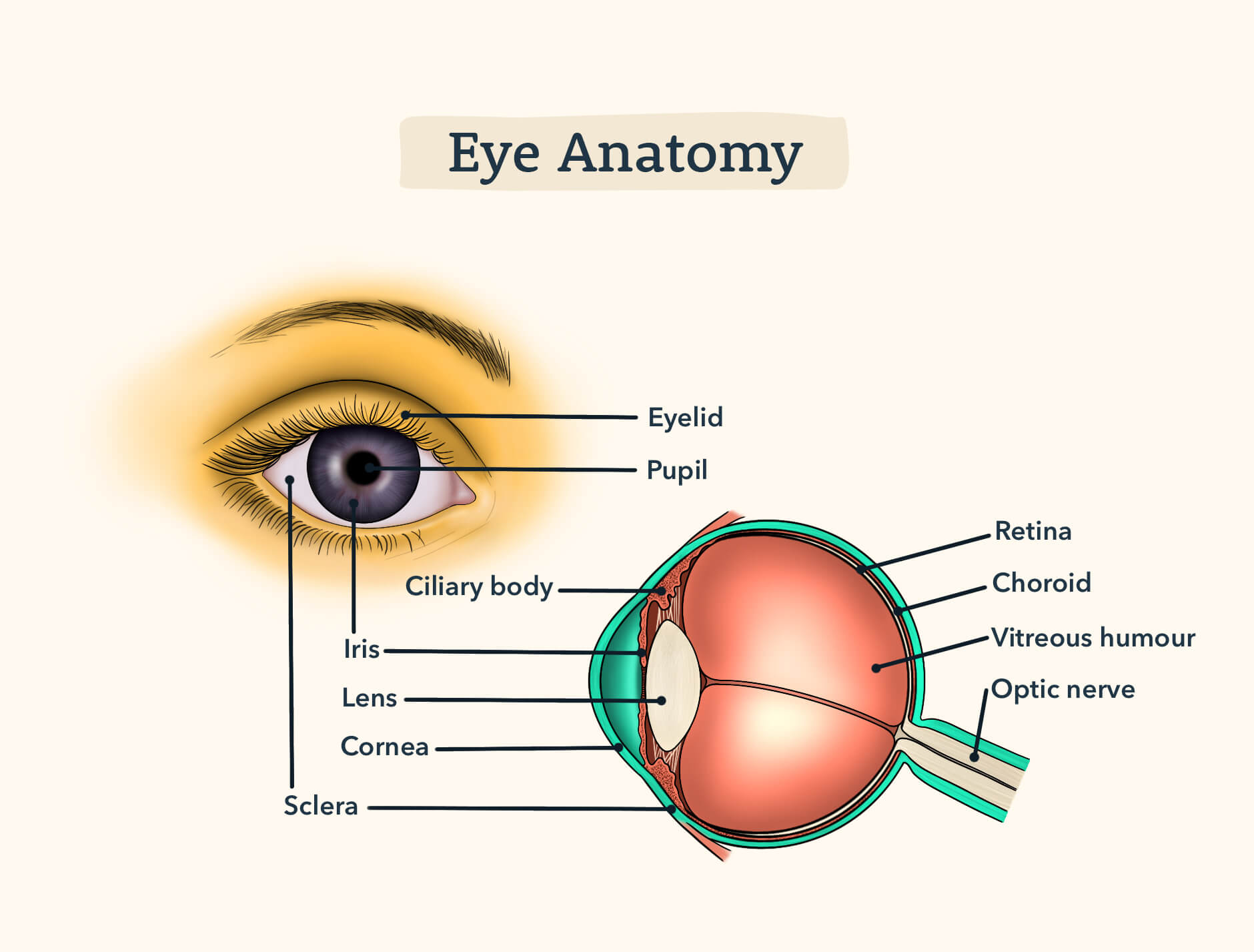
Eyes
- Retina: a layer in the back of the eyeball, where visual images are formed
- Rods: cells that are responsible for seeing in dim, dark lighting
- Cones: cells that are responsible for color, and seeing in bright light
- Iris: this is the color portion of the eye, in the image below it’s a hazel blue
- Vitreous Humor: gel-like fluid that fills the eyeball
- Fovea centralis: a cavity in the eye that holds the cone cells
10. The Reproductive System
You know when your mom or dad talks about how cute you were when you were “little”? I bet you never thought about just how truly tiny you once were! In fact, we all start our lives as just itty bitty cells. From there we grow and grow, until one day, we are fully formed humans preparing to take the HESI! Just think – without the reproductive system, you wouldn’t even be here today!
To prepare for questions regarding the reproductive system, you should begin by memorizing the stages of mitosis as well as the function of the following key terms:
- Fertilization: The union of the egg and the sperm
- Fallopian Tubes: The tubes in which the eggs travel from the ovaries to reach the uterus
- Estrogen: The hormone which regulates the female reproductive system
- Testosterone: The hormone which regulates the male reproductive system; females have it in very small amounts
- Zygote: The result of fertilization when the egg and sperm become one
- Ovaries: Produces hormones and releases one egg each month for fertilization.
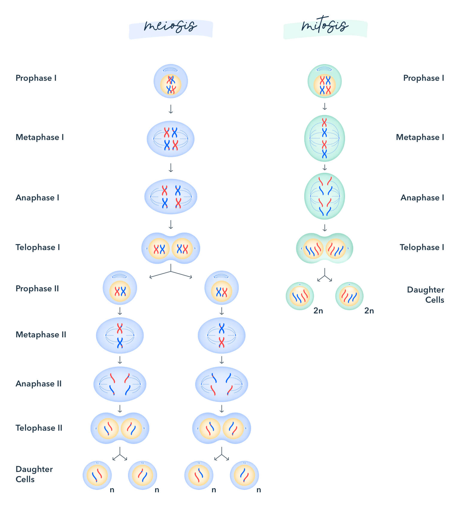
Stages of Mitosis: (note that a useful mnemonic for stages of mitosis is Party More At The Club)
- Prophase: Chromosomes start to form spindle shapes and separate
- Metaphase: The chromosomes start to align into the middle
- Anaphase: Chromosomes are now two separate entities and move away from the middle to the opposite sides
- Telophase: The nuclear membrane begins to pinch itself around each set of chromosomes
- Cytokinesis: The cell splits into two daughter cells
Each cell carries 23 chromosomes.
Score High on the HESI A2 Anatomy and Physiology Section
And, just like that, we wrap up the general topics of anatomy and physiology! We do not want to beat a dead horse over and over, but we must say that it is imperative to remember that knowing the main functions and anatomy of the above mentioned human body systems will help you succeed in the HESI. Having a concrete understanding of how these important systems work will get you the score you are looking for to get into nursing school or other health career programs. There is so much material about the HESI on the internet, but the above review on anatomy and physiology is an informative, pinpointed outline on what to study specifically for the HESI A2 anatomy and physiology section exam.
