


When an atrium tells a lie, what do we call it? A fib!
Have you ever asked for advice and received the answer, “Listen to your heart?” While well-meaning, if you actually took that advice, all you’d hear is lub dub lub dub lub dub…
And though lub dub seems basic, it’s actually incredibly complex and, quite frankly, amazing. In this article, we are delving into the inner workings of the human heart.
Ready? Take notes!
Here’s the big picture: the human heart is the pump that circulates blood through the body. It pumps blood through the arteries, to arterioles, and then to capillaries.
Capillaries are the site of exchange between blood and tissues of the body. What is exchanged?
- Gases
- Nutrients & Electrolytes
- Waste
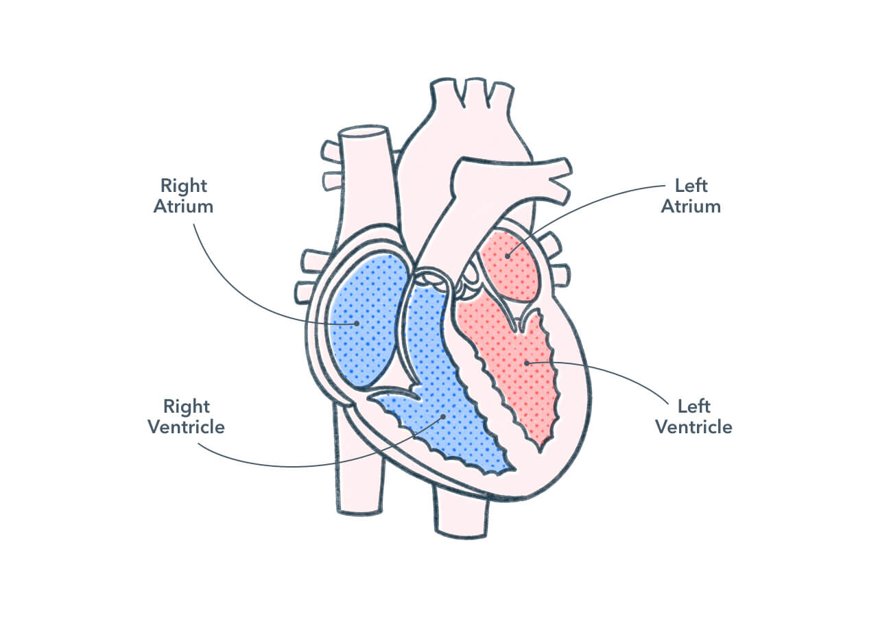
The human heart consists of four-chambers — two atria and two ventricles. The four chambers work in synchronization to contract and relax. During each cardiac cycle (aka heart beat):
- Atria contract/ventricles relax (systole). Then,
- Ventricles contract/atria relax (diastole). Then,
- Both atria and both ventricles relax.
The point of all of this? To change pressure within the different chambers to cause the opening and closing of valves. Valves allow the blood to flow in a unidirectional path through the heart.
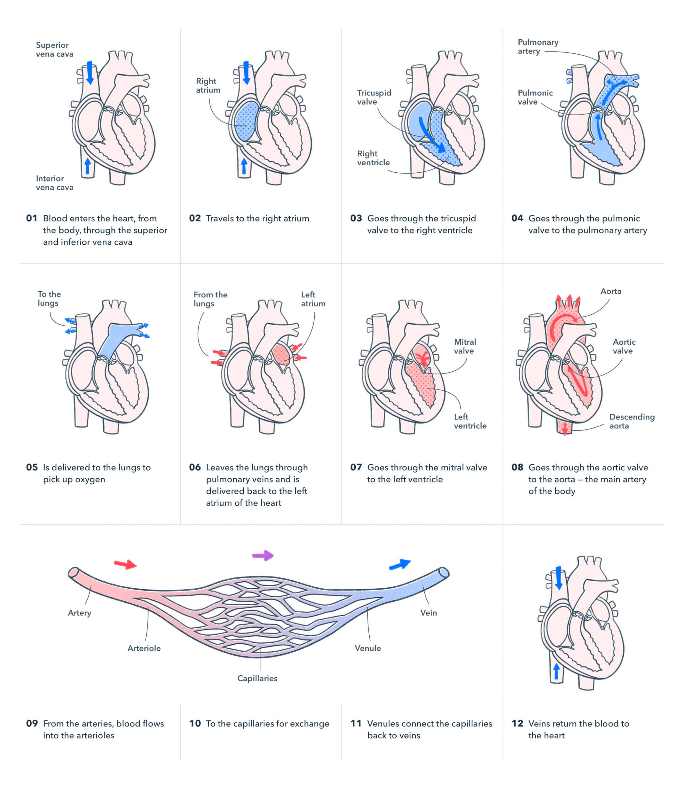
Let’s zoom in from the big picture and examine the path of blood flow
This flow of blood is part of what’s called a “closed circuit.” It’s called closed because the blood is enclosed (within vessels) and circuit because it’s a repeating path.
- Blood enters the heart, from the body, through the superior and inferior vena cava
- And travels to the right atrium
- Then goes through the tricuspid valve to the right ventricle
- And goes through the pulmonic valve to the pulmonary artery
- Then is delivered to the lungs to pick up oxygen
- And leaves the lungs through pulmonary veins and is delivered back to the left atrium of the heart
- Then goes through the mitral valve to the left ventricle
- And then goes through the aortic valve to the aorta—the main artery of the body
- From the arteries, blood flows into the arterioles
- And then to the capillaries for exchange
- Venules connect the capillaries back to veins
- Veins return the blood to the heart
The circulatory system can be divided into two main circuits:
1. Pulmonary circuit
Veins and venules send deoxygenated blood to the lungs for gas exchange (CO2 is dumped and O2 is picked up).
2. Systemic circuit
Arteries and arterioles send oxygenated blood to the body to supply nutrients and remove waste.
But, what actually makes the heart beat? Special cardiac muscle cells work together to conduct electrical impulses throughout the heart. These electrical signals stimulate contraction. These cells are highly concentrated in the sinoatrial node (SA), which is also known as the pacemaker.
Feeling stressed? Perhaps all of this intense studying is raising your blood pressure! And speaking of blood pressure…
Blood pressure is taken as the force against the blood vessels produced by the flow of blood. It is measured as systolic pressure/diastolic pressure using a sphygmomanometer.
Many things affect blood pressure:
- Cardiac output: volume of blood released from the ventricle
- Peripheral resistance: resistance of blood vessels to blood flow (friction), which is influenced by CO2 & O2
- Blood volume and blood viscosity
As blood leaves the heart, it is at its highest pressure. The pressure decreases as the branching of vessels increases. Once blood reaches the capillaries, the pressure is very low. This allows filtration to occur.
Since we’ve discussed the path of blood leaving the heart, let’s now examine the return
After filtration and reabsorption at the capillary level, the blood enters venules, then veins. The movement of blood through veins is made possible by skeletal muscle contraction – remember, the force of contraction is weaker at the venous level than the arterial level.
Making its way back to the heart, blood flow is controlled by valves that open when blood is flowing towards the heart, but moving to a closed position if blood flows backward.
You’ve now examined the steps of one complete cycle of the incredibly complex circulatory system. Congratulations on making it through. Cardiac anatomy and physiology is not for the faint of heart!
